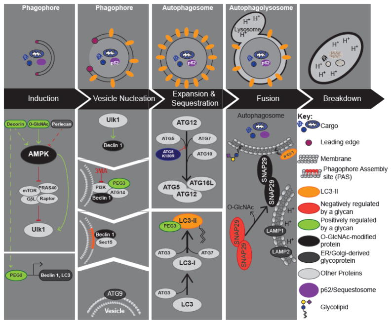
Figure 1. Autophagy.
Upper Panel: Autophagy can be broken down into 5 key steps: 1) Induction; 2) Vesicle nucleation; 3) Expansion and Maturation; 4) Fusion with the Lysosome; and 5) Degradation. Critical regulatory events for each step are shown below. Lower Panel: Pro- and anti-autophagy signals converge on ULK1 (Atg1 in yeast), which performs the first committed step in autophagy. Activation of the ULK1 complex leads to the recruitment of the Beclin 1-VPS34 complex at the PAS (phagophore assembly site). Maturation of the phagophore is promoted by lipids contained in ATG9 vesicles. Expansion and sequestration requires two ubiquitin conjugating systems, which result in the lipidation of LC3 and its targeting to immature autophagosomes (phagophores). Autophagosomes fuse with the lysosome, in a manner dependent on SNAP29.The key proteins involved in regulating each step are shown, and are in light grey unless glycosylated or regulated by a glycan (lower panel). Proteins that are O-GlcNAc modified are indicated in black, whereas other types of glycosylation are highlighted in dark grey. The leading edge is highlighted by purple cicle.

