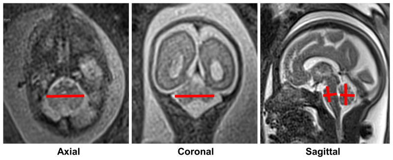
Figure 2. Linear MRI measurements of the fetal posterior fossa.
Examples of measurements taken of the six posterior fossa structures from 2D MR images. Axial and coronal transcerebellar diameter measurements were taken at the point of maximal length (left and middle panel). As is evident in the middle panel, it is common to obtain a slightly out of plane view of the cerebellum in one or more planes, which can introduce error into linear measurements. Vermis and pons heights and anterior-posterior diameters were achieved at the mid-sagittal plane at the level of the corpus callosum. Note that 3D measurements were achieved in an identical fashion.

