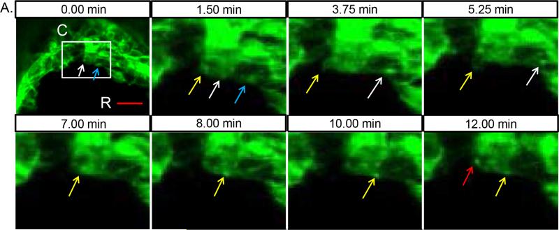
Fig. 4. Membrane shuttling during zipping of the neural folds.
(A) High speed imaging of NNE during hindbrain zipping in a 9 somite embryo. White box indicates region of additional magnification in the remaining images taken over a 12 minute time course. A bright foci of membrane (highlighted by white arrow) moves from the cell at the midline to the neighbouring cell rostral along a filopodial connection. At the start of the movie, a membrane focus is already traversing along the filopodial extension between the two cells (blue arrow). Between 3.75 min and 5.25 min, a membrane bleb (highlighted by yellow arrow) is pulled back into the midline cell followed by the formation of a second focus of membrane that then moves into the next cell. At 12 min, an additional membrane focus is initiated (red arrow). Scale bar = 20μm. R=rostral, C=caudal

