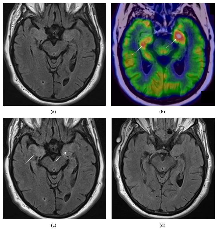
Figure 1.
Neuroimaging of VGKC antibody. The initial, unremarkable axial T2/FLAIR MRI of the brain (a) followed by FDG-PET showing significant asymmetric hypermetabolism of the bilateral mesial temporal lobes (arrows; (b)) with repeat axial T2/FLAIR demonstrating hyperintensity in bilateral mesial temporal lobes (arrows; (c)) that significantly improved by the last T2/FLAIR (d).

