
Fig. 1.
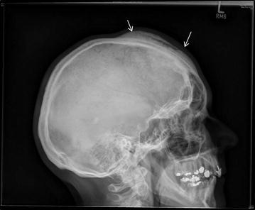
Lateral X-ray of the skull shows an extensive osseous lesion of the diploe with periosteal new bone formation (arrows) along the external table of the anterior skull
Official websites use .gov
A
.gov website belongs to an official
government organization in the United States.
Secure .gov websites use HTTPS
A lock (
) or https:// means you've safely
connected to the .gov website. Share sensitive
information only on official, secure websites.
Lateral X-ray of the skull shows an extensive osseous lesion of the diploe with periosteal new bone formation (arrows) along the external table of the anterior skull