
Fig. 4.
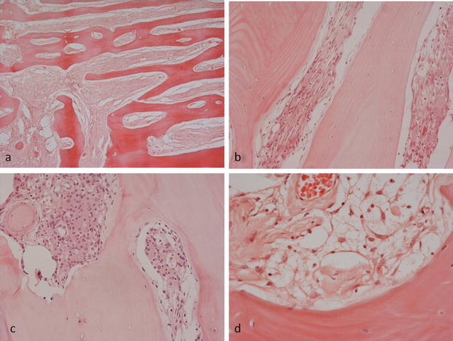
Histopathological features of PIM showing a extensive organised reactive bone formation within the lesion. Intertrabecular fibrous tissue contains b a proliferation of cells with spindle-shaped and vesicular nuclei; c scattered meningotheliomatous whorled collections of cells; d occasional cells with clear, vacuolated cytoplasm