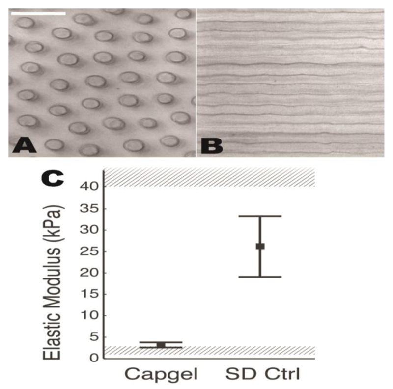
Figure 1.

Image series of Capgel and cells cultured within the scaffold at different time points. (A,B) Phase-contrast images perpendicular (A) and parallel (B) to channel long axis. (C) Mechanical properties of Capgel are above baseline modulus shown to support cardiomyocyte function (1–3 kPa, lower shaded region,(Bhana et al., 2010; Engler et al., 2008; Jacot et al., 2008; Bajaj et al., 2010)and below fibrotic modulus shown to induce dysfunction (40–70 kPa, upper shaded region, Bhana et al., 2010; Engler et al., 2008; Jacot et al., 2008). Mean and standard deviation for polymerized Capgel (n = 49 indentations) and 12-week-old Sprague Dawley normal ventricular myocardium (16 indentations) are shown.
