Figure 5.

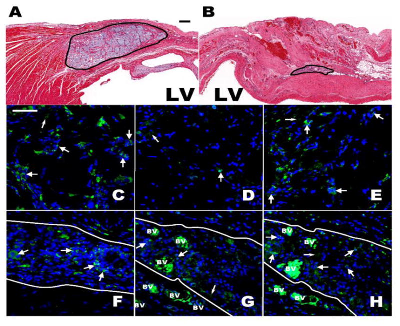
Histological and Immunofluorescent assesment 4 and 8 weeks post Capgel injection into the antero-septal LV Wall. (A,B) Brightfield micrograph of H&E stained antero-septal LV wall showing the Capgel area after 4 (A) and 8 (B) weeks; Scale bar = 200 μm for both. (C–H) Immunofluorescent staining (green) of macrophages populating the Capgel area 4 (C–E) and 8 (F–H) weeks post gel injection: CD68 (C,F), CD86 (D,G) and CD206 (E,H). Capgel area is between solid white lines (F–H). Large white arrow heads point to positively stained cells/clusters of cells for a given antibody; small white arrow heads point to nonspecific staining and/or autoflourescences of the injected Capgel. Blue = DAPI stained cell nuclei and BV = blood vessel; note the intense autofluorescence of RBCs within the vessels. Scale bar = 50 μm for all.
