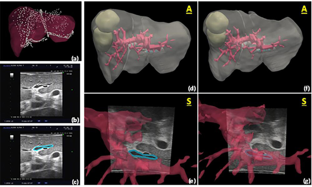
Figure 3.
Using methods from Rucker et al.92, (a) preoperative liver model and registered swabbed point cloud acquired from intraoperative liver surface are shown with ultrasound image of segment III portal pedicle in (b), and (c) showing manual segmentation of structure. In (d) we see the planned model cloud with ultrasound slice (white arrow) showing alignment of pedicle based on rigid registration, and (e) is the close-up. Notice how inferior vessel region does not align with corresponding vascular structure. In (f), a deformation correction driven by the closest point mismatch in data shown in (a) has been performed, and (g) shows the new location of the structure as well as modified shapes to vasculature. It should be noted that in this example ultrasound data was used for validation only, i.e. no localized ultrasound structures were used within the alignment algorithm, only data shown in (a) was used. We should further note that the alignment error of this feature after rigid registration was estimated at 5.5 +/− 2.6 mm (10.9 mm maximum error). The error was reduced to 2.4 +/− 1.5 mm (5.4 mm maximum error) after model-based deformation correction.

