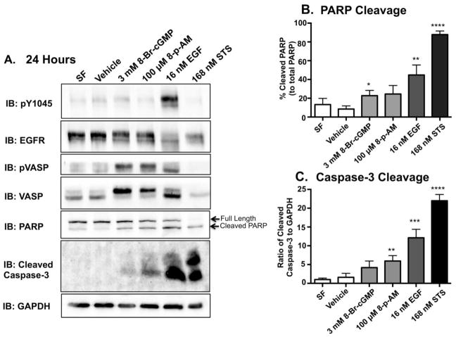
Figure 4. PKG agonists induce apoptosis in the MDA-MB-468 cell line.
A. MDA-MB-468 cells were serum starved overnight. With the exception of Staurosproine (STS), the cells were then subjected to each experimental condition for 24 hours. MDA-MB-468 cells were treated with STS for 3 hours prior to being harvested. This was done in order to induce a robust response without inducing catastrophic damage to the cell. The induction of apoptosis was determined by using PARP and Cleaved Caspase-3 antibodies. With the exception of STS, 40 μg of protein per sample were then resolved on a 15% SDS-PAGE. Fifteen μg of protein from the STS sample were resolved on the 15% SDS-PAGE. This was done in order to to ensure that our positive control was within the dynamic range. Quantification of western blot data from cleaved PARP (B.) and Caspase-3 (C.) from three independent experiments. Band intensities were determined using Image J software and plotted relative to total PARP (% cleaved PARP) or GAPDH (ratio of cleaved Caspase-3/GAPDH), respectively. Data are plotted as the average ± SEM (n=3)

