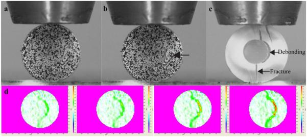
Figure 4.
A dentin-composite disk subjected to diametral compression. a) Disk specimen before loading - its surface had been sprayed with white paint and black powder to create speckles for DIC analysis. b) The same specimen after fracture, with debonding between the restorative material and the dentin ring as indicated by the arrow. c) A different specimen with painting removed to reveal the fracture pattern. d) DIC results showing the emergence and spread of strain concentrations during diametral compression.

