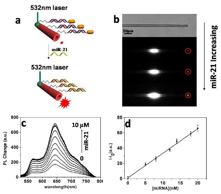
Figure 3.

(a) Schematic illumination of single Au@PDA microtube waveguide system for miRNA-21 detection. (b) White-field image of the single PDA mictrotube and the images of single Au@PDA microtube waveguide sensor upon gradual addition of miR-21. (c) Changes in the out-coupled emission at the tip of PDA microtube with increasing concentrations of miRNA-21. (d) Plot of fluorescence-enhancement of the out-coupled tip emission of PDA microtubes upon gradual addition of miR-21 (ranged from 0 to 20nM). I0 and I represent the tip emission intensity of PDA microtube at 640 nm before and after displacement reaction with miRNA-21, respectively.
