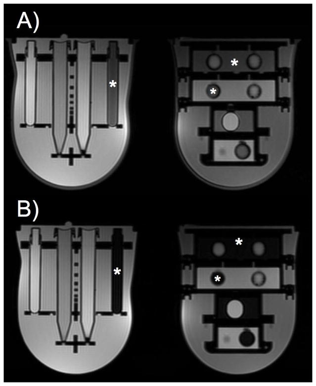
Figure 3.
Axial T2-weighted images acquired without (A) and with (B) spectral fat suppression demonstrate that fat signal is suppressed in the adipose tissue mimic (locations of adipose tissue mimic noted with *). Additionally, signal from the silicone shell, at the boundary of the breast phantom, is suppressed using spectral suppression techniques (B).

