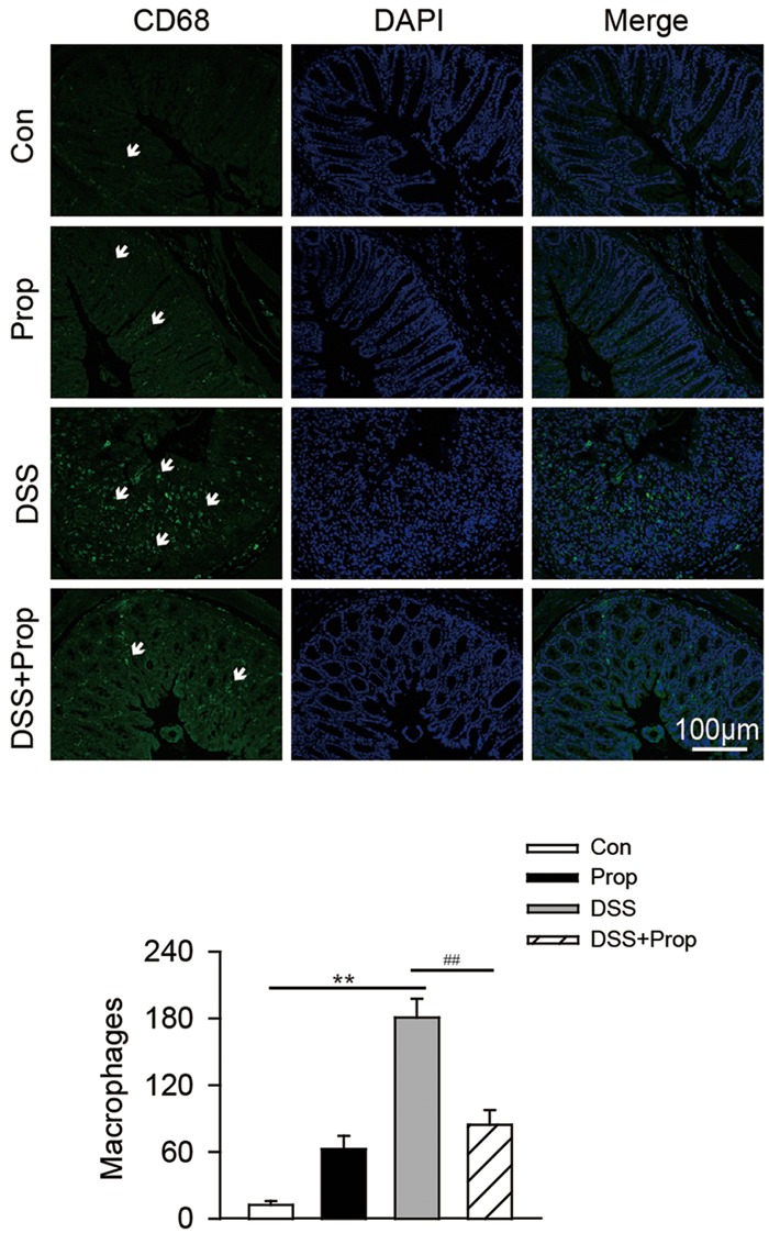
FIGURE 5.

The effects of sodium propionate on macrophages with CD68 marker in the colonic tissue. Macrophages were determined by immunofluorescence in colonic tissues. CD68 was used as the marker of macrophage/monocyte infiltration in the colon of mice and visualized by fluorescence microcopy (green staining). Nuclei were stained with DAPI (blue staining). Arrows indicate CD68-labeled macrophages in the colon. Images were representative of 6–8 mice in each group. Scar bars were 100 μm. Morphometric analysis of macrophages in the colon was also performed. Data are presented as mean ± SD. n = 6–8 per group, ∗P < 0.05, ∗∗P < 0.01 vs. control group; #P < 0.05, ##P < 0.01 vs. DSS group.
