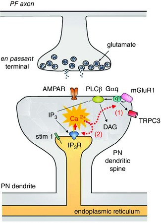
Figure 2. Schematic diagram of the mGluR–Ca2+ hypothesis .

Glutamate release from PFs activates both AMPARs and mGluR1 GPCRs. As indicated by the black arrows, mGluR1 is coupled to PLCβ, which leads to release of Ca2+ from IP3Rs on the endoplasmic reticulum. Ca2+ exerts positive feedback on mGluR1 transduction at a step early in the cascade (1) as well as at the IP3R (2). Thus, elevations in Ca2+ will exacerbate the IP3R hyperactivity observed in SCA2. Modified from Hartmann et al. (2011). DAG, diacylglycerol.
