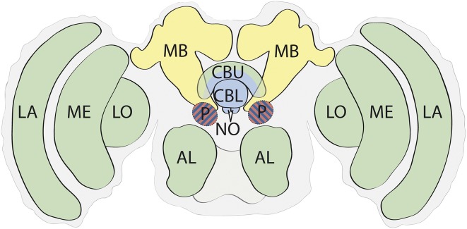
Fig. 2.
Basic functional anatomy of the insect brain (not to scale). The structures of the insect brain create an integrated neural model of the state of the insect in space that is functionally analogous to that described for the vertebrate brain in Fig. 1. Regions are colored to reflect the major functions described in Fig. 1A. Vision and smell are primarily processed by dedicated sensory lobes, which function to refine and enhance sensory representations and enhance distinctions between similar stimuli (95, 141). Primary odor processing is performed in the antennal lobes (AL). Visual processing is performed by the lamina (LA), medulla (ME), and lobula (LO). The MB supports learning and memory (142–146). The CX is anatomically variable between insect orders but typically is composed of the central body upper (CBU), central body lower (CBL), and noduli (NO). It has several specializations for processing spatial information corrected for self movement (75, 76, 83, 87). The protocerebrum (P) is an anatomically complicated region. Modulatory and inhibitory connections to and within the protocerebrum convey information on physiological state (94, 95), and structures within the protocerebrum, particularly the lateral accessory lobe, are involved in integration of information, hence the hatched shading.

