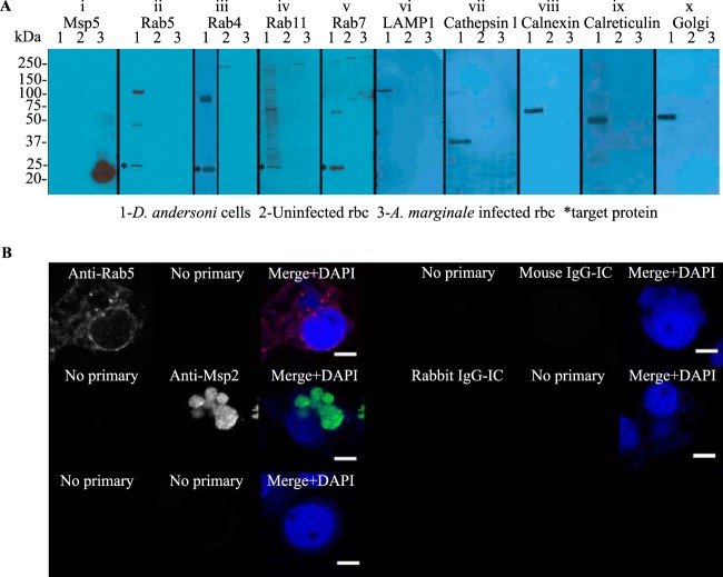
FIG 1.
Western blot detection of endosomal and secretory pathway proteins from D. andersoni and specificity of immunofluorescence assays. (A) Equivalent amounts of protein (50 μg) from DAE100T cells (lane 1), uninfected erythrocytes (lane 2), and A. marginale-infected erythrocytes (lane 3) are in each lane. In panel i, the anti-Msp5 antibody detects a protein of the expected size (20 kDa), confirming the presence of A. marginale in the infected erythrocytes. In panels ii to x, the anti-Rab and anti-organelle antibodies detect bands of the expected size in DAE100T cells (lane 1) as follows: Rab5 (26 kDa), Rab4 (23 kDa), Rab11 (24 kDa), Rab7 (23 kDa), LAMP1 (120 kDa), cathepsin L (37 kDa), calnexin (67 kDa), calreticulin (50 kDa), and 58K Golgi protein, as indicated by the asterisks in panels ii, iii, iv, and v. None of the antibodies recognize A. marginale (lane 3). (B) To demonstrate the specificity of the primary and secondary antibodies, A. marginale-infected DAE100T cells were incubated with only secondary antibodies (no primary) or primary antibodies targeting cellular markers (anti-Rab5 antibody shown here), A. marginale Msp2, or murine or rabbit isotype controls (IgG-IC). All assays were performed using anti-rabbit IgG (red) and anti-murine IgG (green) secondary antibodies.

