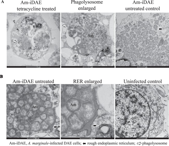
FIG 11.
The AmVs are associated with the rough endoplasmic reticulum and mature to phagolysosomes in the absence of bacterial protein synthesis. D. andersoni cells that were infected with A. marginale were evaluated by transmission electron microscopy. (A) In tetracycline-treated infected D. andersoni cells (tetracycline treated, enlarged), colonies of A. marginale are often within multimembranous phagolysosomes with other organelles (white arrow). An untreated control (Am-iDAE untreated control) demonstrates AmVs unfused to other cellular vesicles and closely associated with recruited RER (black arrow). (B) The rough endoplasmic reticulum is closely applied to the outer aspect of the AmV and is devoid of ribosomes on the contact side (black arrow) in Am-iDAE untreated and at a higher magnification (RER enlarged). Long strands of RER are adjacent to the nucleus in the uninfected control.

