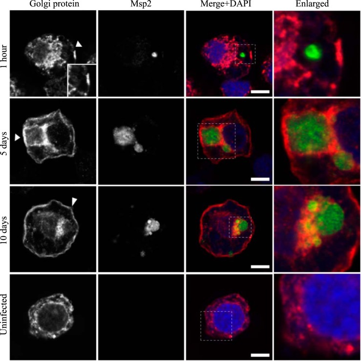
FIG 14.
AmVs accumulate the 58K Golgi protein and retain it throughout infection. Indirect immunofluorescence localization of Golgi proteins using an anti-58K Golgi protein antibody (red) in D. andersoni cells that were synchronously infected with A. marginale labeled with anti-Msp2 antibodies (green) or mock infected with medium (uninfected). AmVs associate with the Golgi marker by 5 days postinoculation, and it is retained at 10 days postinfection. The uninfected control shows the pattern of staining of the 58K Golgi protein in D. andersoni cells. The box in the Merge+DAPI image outlines the area of magnification in the Enlarged column. Scale bars represent 10 μm on a 63×/1.20 objective. The white arrowhead in the first column indicates the location of the AmV and the area enlarged in the inset. All images are at the same magnification, with the exception of the insets and last column, which are enlarged 3-fold.

