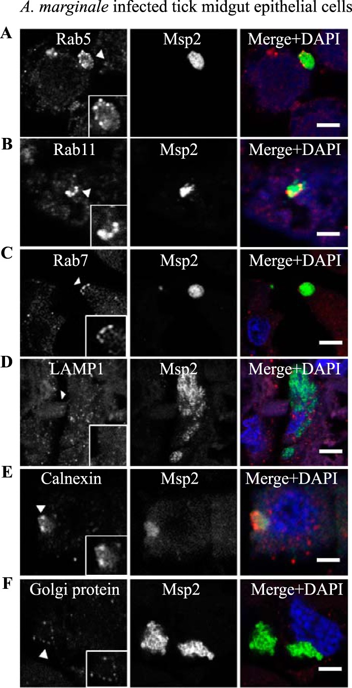
FIG 15.

Aberrant labeling of AmVs with early, recycling, and late endosome markers and association with secretory markers in D. andersoni in vivo. The characteristics of the AmVs were evaluated in tissue sections from A. marginale (green)-infected D. andersoni midguts. AmVs in the tick midgut recruited the early endosome marker Rab5 (A), the recycling endosome marker Rab11 (B), and the late endosome marker Rab7 (C). The AmVs did not associate with the lysosome marker LAMP1 (D). The endoplasmic reticulum marker calnexin (E) and the Golgi marker 58K Golgi protein (F) associated with the AmVs. All cellular markers are in red. Scale bars represent 10 μm on a 63×/1.20 objective.
