FIG 7.
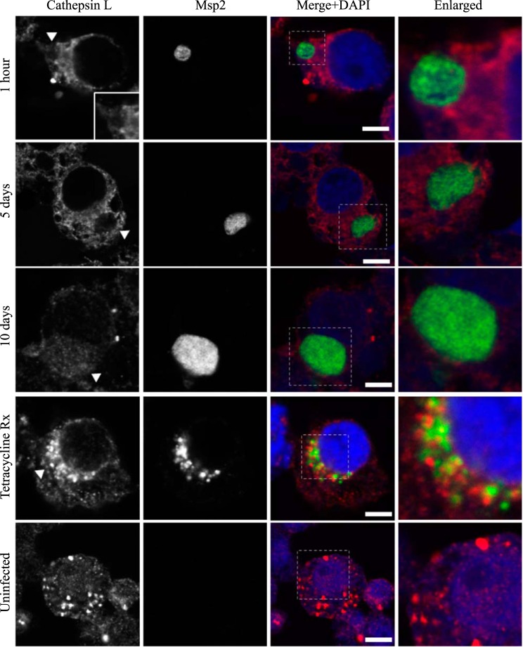
AmVs do not accumulate the lysosome protease cathepsin L throughout infection. Indirect immunofluorescence localization of cathepsin L using anti-cathepsin L antibodies (red) in D. andersoni cells that were synchronously infected with A. marginale labeled with anti-Msp2 antibodies (green) or mock infected with medium (uninfected). Labeling with cathepsin L in AmVs is not apparent at 1 h, 5 days, and 10 days postinoculation. Inhibition of bacterial protein synthesis (Tetracycline Rx) resulted in cathepsin L labeling within the majority of AmVs. The uninfected control shows the pattern of staining of cathepsin L in D. andersoni cells. The box in the Merge+DAPI image outlines the area of magnification in the Enlarged column. Scale bars represent 10 μm on a 63×/1.20 objective. All images are at the same magnification, with the exception of the insets and last column, which are enlarged 3-fold. The white arrowhead in the first column indicates the location of the AmV and the area enlarged in the inset.

