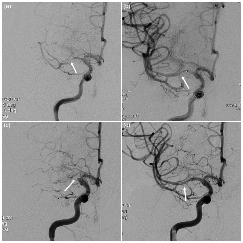
Figure 2.
A high-grade right MCA stenosis. (a) Pre-stenting right ICA angiogram reveals tight stenosis (99%) of M1 segment. (b) First-time post-stenting right ICA angiogram reveals resolution of the critical stenosis. (c) The follow-up (3 days) right ICA angiogram shows complete in-stent thrombosis. (d) After thrombolytic recanalization and balloon-expandable Helistent (2.5 × 7 mm) deployment, right ICA angiogram shows patent flow with some minimal filling defects representing thrombotic debris.

