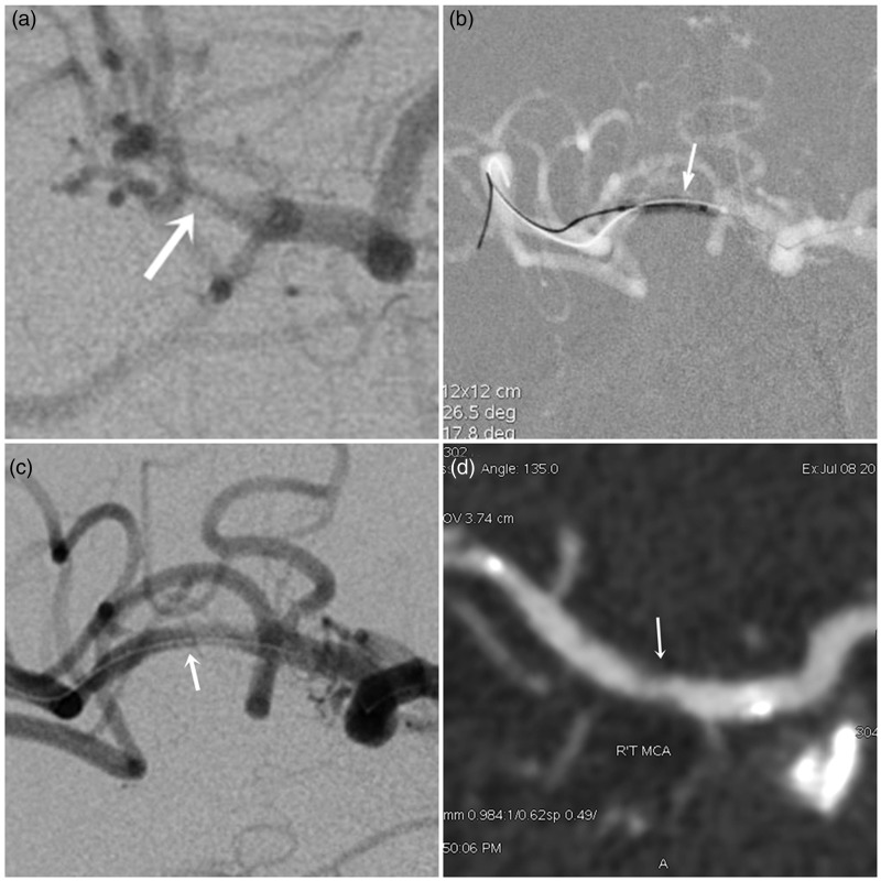
Figure 4.
A high-grade right MCA stenosis. (a) Pre-stenting right ICA angiogram shows right proximal M2 stenosis (90%). (b) Undersized PTA was performed by using a Rafale balloon (1.5 × 10 mm) for the M2 stenotic lesion. (c) Post-stenting angiogram after Enterprise stent (4.5 × 22 mm) deployment shows less than 10% diameter stenosis of right M2 segment. (d) Follow-up CTA 18 months after PTAS demonstrates in-stent restenosis with nearly 55% luminal narrowing.

