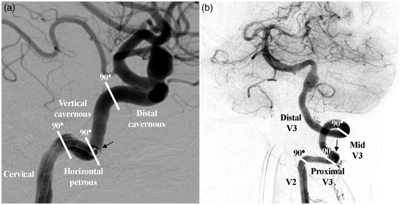
Figure 1.
Lateral view of the internal carotid artery (a) and anteroposterior view of the vertebral artery (b) illustrating the system used to assess 90° turns and landing and final position. Panel (a) shows the Benchmark intracranial guide catheter landing position in the horizontal petrous segment of the internal carotid artery during Pipeline embolization of a paraophthalmic internal carotid artery segment aneurysm. Panel (b) shows the landing position of the Benchmark in the proximal V3 segment of the vertebral artery during Pipeline embolization of a fusiform aneurysm involving the origin of the posterior inferior cerebellar artery.

