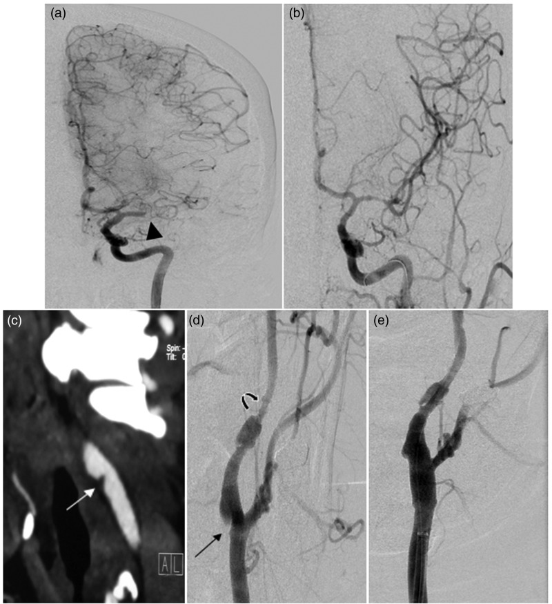
Figure 2.
(a) Anteroposterior (AP) left ICA DSA demonstrates occluded left MCA at the mid-M1 segment (arrow head) with occlusion of the lateral lenticulostriate arteries, but robust collaterals from the left ACA. (b) AP left ICA DSA after mechanical thrombectomy with stent retrieval demonstrates successful recanalization of the left MCA and its inferior division, and slow antegrade flow into the superior division (TICI = 2b). (c) Reformatted left oblique CTA reveals a focal stenosis at the origin of the left ICA secondary to a shelf-like hypodense projection (white arrow), raising the possibility of carotid web or noncalcified atheroma. (d) Left CCA anterior oblique DSA confirms a sharp-edged triangular ridge on the posterolateral aspect of the left ICA (black arrow) consistent with a carotid web and a separate downstream segmental stenosis with pseudoaneurysm consistent with an iatrogenic dissection. (e) Left anterior oblique CCA DSA post-treatment demonstrates the Xact stent (black arrow) deployment across the left ICA origin and the Neuroform stent reconstruction and angioplasty across the iatrogenic dissection (arrow heads) with no residual stenosis. ICA: internal carotid artery; DSA: digital subtraction angiography; MCA: middle cerebral artery; ACA: anterior cerebral artery; TICI: Thrombolysis in Cerebral Ischemia score; CCA: common carotid artery.

