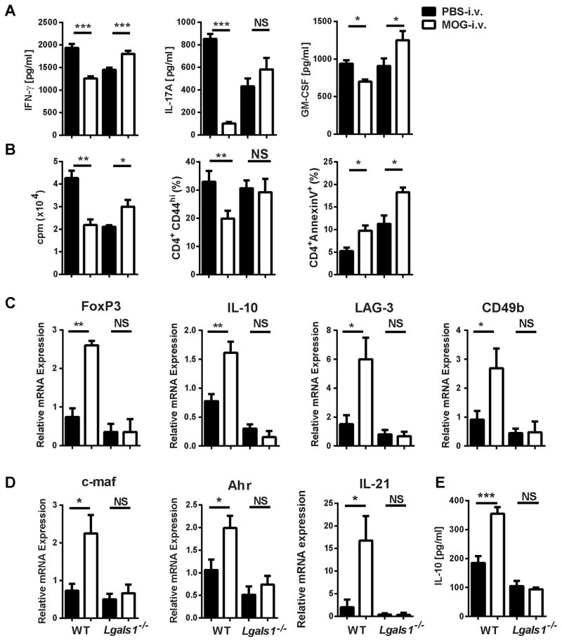
Figure 4. Lack of galectin-1 leads to increased pro-inflammatory responses and reduced numbers of Tregs in the periphery of i.v. tolerized mice.
Splenocytes from MOG-i.v. and PBS-i.v. EAE WT and Lgals1−/− mice on day 25 p.i. were stimulated for three days with MOG35-55. (A) Cell culture supernatants were assayed for IFN-γ, IL-17A and GM-CSF by ELISA (B) Proliferation was measured in splenocytes after three days of culture with MOG35-55. Tritiated adenine was added after 54 hours of culture; its incorporation was measured after 18 hours. CD4+AnnexinV+ and CD4+CD44hi T cells from the spleen were analyzed by flow cytometry on day 25 p.i. (C) IL-10, LAG-3, CD49b and FoxP3 mRNAs in splenocytes were measured using RT-PCR. (D) IL-10 concentrations in cell culture supernatants were measured by ELISA. (E) c-maf, Ahr and IL-21 mRNAs in splenocytes were measured using RT-PCR. Data shown are one representative of four experiments. Experiments show mean ± SD. *, p<0.05, **, p < 0.01, ***, p < 0.001, unpaired Student’s t test.

