Figure 3.
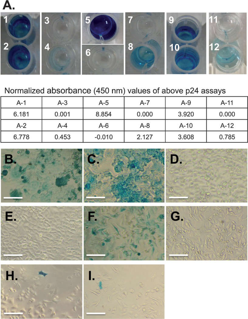
Control HIV-1 replication with Amber suppression. A) p24 assay after transfection of 293T with pSUMA variants. The absorbance values were determined at 450 nm after the colorimetric reactions were stopped by the addition of 1m H2SO4. The well 11 was used as blank for all measurements. 1, wild-type pSUMA only; 2, wild-type pSUMA + tRNATyr–AzFRS pair + 1 mm AzF; 3, pSUMA-Tyr132 + tRNATyr–AzFRS pair, without AzF; 4, pSUMA-Tyr132 + tRNATyr–AzFRS pair, with 1 mm AzF; 5, pSUMA-Tyr132 + tRNATyr–TyrRS pair; 6, pSUMA-Tyr132; 7, pSUMA-Ala119 + tRNATyr–AzFRS pair, without AzF; 8, pSUMA-Ala119 + tRNATyr-AzFRS pair, with 1 mm AzF; 9, pSUMA-Leu365 + tRNATyr-AzFRS pair, without AzF; 10, pSUMA-Leu365+tRNATyr-AzFRS pair, with 1 mm AzF; 11, negative ELISA control, no p24; 12, positive ELISA control, 125 pgmL−1 p24; B)–I) Infection assays with TZM-bl cells: B) infected with virus collected from (A-1); C) infected with virus collected from (A-2); D) infected with virus collected from (A-4); E) infected with virus collected from (A-3); F) infected with virus collected from (A-5); G) infected with virus collected from (A-6); H) infected with virus collected from (A-8); I) infected with virus collected from (A-10). Scale bars, 100 µm.

