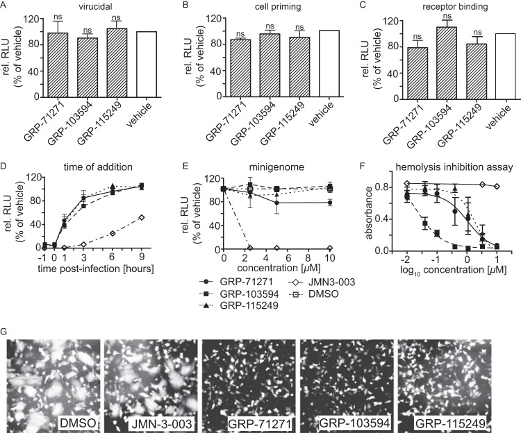
FIG 3.
Mechanistic characterization of selected hits. (A to C) Pretreatment of virus or cells with compounds. IAV-WSN-nanoLuc (A) or BEAS-2B cells (B) were preincubated with 10 μM compound for 2 h, or the virus was adsorbed to cells for 2 h at 4°C in the presence of compound (C). After compound removal and, for panels A and B, infection of cells, luciferase activity was determined after incubation at 37°C for 30 h. Bars represent averages for three independent experiments and SD; one-way ANOVA with Sidak's multiple-comparison posttest was used for statistical analysis (ns, not significant [P ≥ 0.05]). (D) Time-of-addition study. Compounds (10 μM final concentration) or an equivalent volume of vehicle (DMSO) was added at the specified times postinfection to BEAS-2B cells infected with IAV-WSN-nanoLuc. Luciferase activities in all samples were determined after incubation at 37°C for 20 h. Values represent averages for three independent experiments ± SD. (E) IAV-WSN-based minireplicon assay. 293T cells transiently expressing IAV-WSN NA, NP, PB1, and PB2 proteins and transfected with an IAV-WSN-based minigenome reporter containing firefly luciferase (31) were incubated in the presence of different compound concentrations or an equivalent volume of vehicle (DMSO). Luciferase activity was determined after incubation at 37°C for 36 h. Values represent averages for three independent experiments ± SD. (F) Hemolysis inhibition assay. Chicken red blood cells were mixed with purified IAV-WSN and compounds or an equivalent volume of vehicle (DMSO), subjected to pH 5.1, and incubated for 30 min at 37°C. The absorbance of released hemoglobin was measured at 410 nm and corrected for baseline signals of equally treated but mock-infected cells. Values represent averages for three independent experiments ± SD. (G) Cell-to-cell fusion assay. BSR T7/5 cells were transfected with expression plasmids encoding IAV-WSN HA or EGFP at a relative ratio of 10:1, followed by trypsin treatment and exposure to pH 5.1 for 5 min at 37°C in the presence of 10 μM compound or an equivalent volume of vehicle (DMSO). Syncytium formation was assessed microscopically after 30 min of incubation at 37°C. Representative fields of view are shown. Magnification, ×100.

