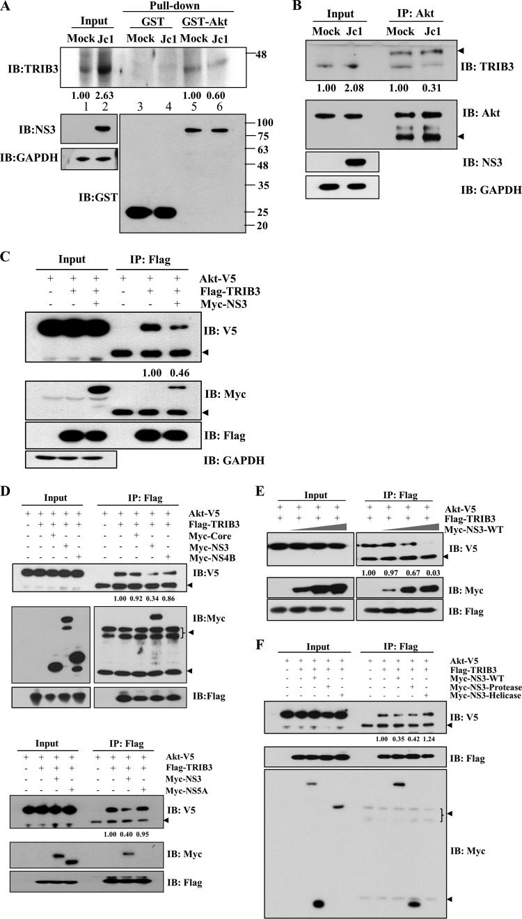
FIG 6.
HCV NS3 interrupts the interaction between TRIB3 and Akt. (A) Huh7 cells were either mock infected or infected with Jc1. At 6 days postinfection, total cell lysates were harvested and incubated with either GST- or GST-Akt-conjugated glutathione beads. Bound proteins were analyzed by immunoblot analysis using an anti-TRIB3 antibody. Protein band intensities of input TRIB3/GAPDH (lanes 1 and 2) and pulled-down TRIB3 normalized by Jc1/mock (lane 6 versus lane 5) were analyzed by using ImageJ. (B) Huh7 cells were either mock infected or infected with Jc1. At 6 days postinfection, total cell lysates were immunoprecipitated with an anti-Akt antibody, and bound protein was then immunoblotted with an anti-TRIB3 antibody. The arrowheads indicate IgG. Protein band intensities of input TRIB3/GAPDH (lanes 1 and 2) and immunoprecipitated TRIB3 normalized by Jc1/mock (lane 4 versus lane 3) were determined by using ImageJ. (C) HEK293T cells were cotransfected with V5-tagged Akt and vector- or Flag-tagged TRIB3 in the absence or presence of Myc-tagged NS3. Thirty-six hours after transfection, cell lysates were immunoprecipitated with an anti-Flag antibody, and bound proteins were analyzed by immunoblot analysis using either an anti-V5 or an anti-Myc antibody. The arrowheads denote IgG. Protein band intensities of normalized Akt-V5 were analyzed by using ImageJ. (D, top) HEK293T cells were cotransfected with V5-tagged Akt and Flag-tagged TRIB3 together with Myc-tagged core, NS3, and NS4B. Twenty-four hours after transfection, total cell lysates were immunoprecipitated with an anti-Flag antibody, and bound proteins were analyzed by immunoblot analysis using either an anti-V5 antibody or an anti-Myc antibody. The arrowheads indicate IgG. (Bottom) HEK293T cells were cotransfected with V5-tagged Akt and Flag-tagged TRIB3 together with Myc-tagged NS3 and NS5A. Total cell lysates were immunoprecipitated as described above. The arrowhead indicates IgG. Protein band intensities of normalized Akt-V5 were analyzed by using ImageJ. (E) HEK293T cells were cotransfected with Flag-tagged TRIB3 and V5-tagged Akt with increasing amounts (1, 3, and 5 μg) of Myc-tagged NS3. Total cell lysates harvested 36 h after cotransfection were immunoprecipitated with an anti-Flag antibody, and bound proteins were analyzed by immunoblot analysis using either an anti-V5 or an anti-Myc antibody. The arrowhead denotes IgG. Protein band intensities of normalized Akt-V5 were analyzed by using ImageJ. (F) HEK293T cells were cotransfected with V5-tagged Akt and Flag-tagged TRIB3 in the absence or presence of various mutant NS3 constructs. Total cell lysates harvested 36 h after cotransfection were immunoprecipitated with an anti-Flag antibody, and bound protein was analyzed by immunoblot analysis using an anti-V5 antibody. The arrowheads indicate IgG. Protein band intensities of normalized Akt-V5 were analyzed by using ImageJ.

