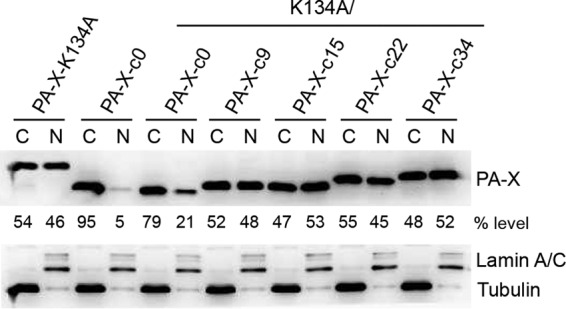
FIG 3.

Subcellular localization of Cal PA-X protein. 293T cells were transfected with the indicated genes in pCAGGS. After 24 h, the cells were fractionated into the nuclear and cytoplasmic materials followed by Western blotting. Expression of PA-X protein was detected using anti-PA-X antibody. Expression of lamin A/C and tubulin was detected as a nuclear (N) and cytoplasmic (C) marker, respectively. The ratios of PA-X in the cytoplasm and nucleus are also shown. The data shown are representative of two independent experiments.
