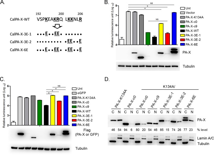
FIG 7.
Role of the 6 basic residues in the PA-X C-terminal region in shutoff activity and localization. (A) Mutations in the PA-X C-terminal region created in this study. The 6 basic residues are shown in boldface and are underlined. Cal PA-X-3E-1 and Cal PA-X-3E-2 contain K195E/K198E/R199E and K202E/K203E/R206E, respectively. Cal PA-X-6E contains all 6 mutations. (B) Luciferase expression from pCAGGS-Luc in 293T cells cotransfected with either empty pCAGGS plasmid (Vector) or the indicated genes in pCAGGS for 24 h. The results are shown as means plus standard deviations (n = 3). Asterisks indicate statistically significant differences (*, P < 0.05 by one-way ANOVA followed by Tukey's multiple-comparison test). NS, not significant (P > 0.05). Expression of PA-X and tubulin was determined by Western blotting as described for Fig. 2B. (C) Luciferase expression from mRNAs in 293T cells cotransfected with the indicated mRNAs for 24 h. The results are shown as means plus standard deviations (n = 3). Asterisks indicate statistically significant differences (*, P < 0.05 by one-way ANOVA followed by Tukey's multiple-comparison test). NS, not significant (P > 0.05). Expression of PA-X, eGFP, or tubulin was determined by Western blotting as described for Fig. 1B. (D) Localization of the indicated PA-X proteins in nuclear and cytoplasmic fractions determined by Western blotting as described for Fig. 3. The data shown are representative of two independent experiments.

