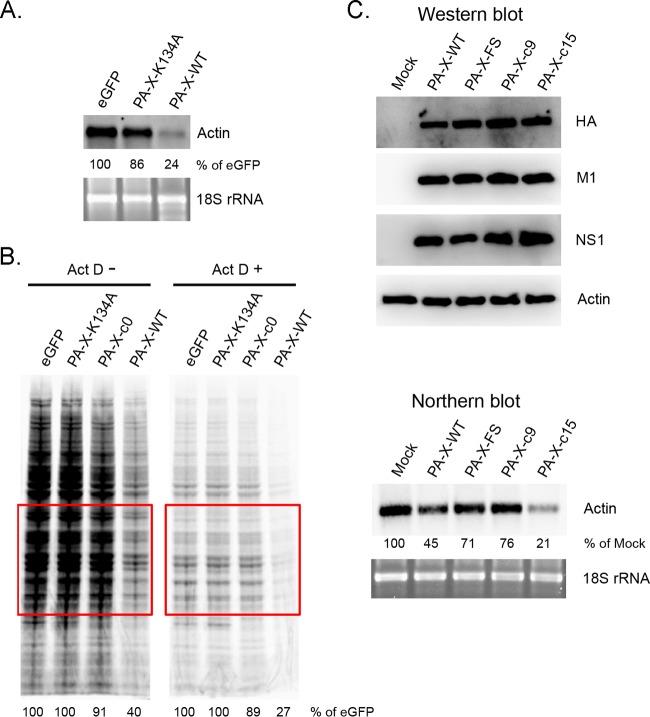
FIG 8.
Effect of PA-X expression on actin mRNA levels and host protein synthesis. (A) 293T cells were transfected with the indicated mRNAs for 24 h. The extracted total RNA was subjected to Northern blot analysis using a DIG-labeled antisense human β-actin probe. The intensities of actin mRNA in each sample were normalized by the intensities of 18S rRNA. The relative level of eGFP was set to 100%. The data shown are representative of two independent experiments. (B) 293T cells were transfected with the indicated mRNAs for 1 h, followed by incubation in the absence or presence of ActD. After 8.5 h of incubation, the cells were labeled with [35S]Met/Cys for 30 min and the lysates were resolved by SDS-PAGE. The intensities of proteins in the indicated area were quantified and normalized to that of control cells expressing eGFP. (C) A549 cells were either left uninfected or were infected with Cal viruses at an MOI of 1.5. At 12 hpi, HA, M1, and NS1 proteins in total cell lysate were detected by Western blotting using specific antibodies. The total RNA was extracted and subjected to Northern blot analysis as described for panel A.

