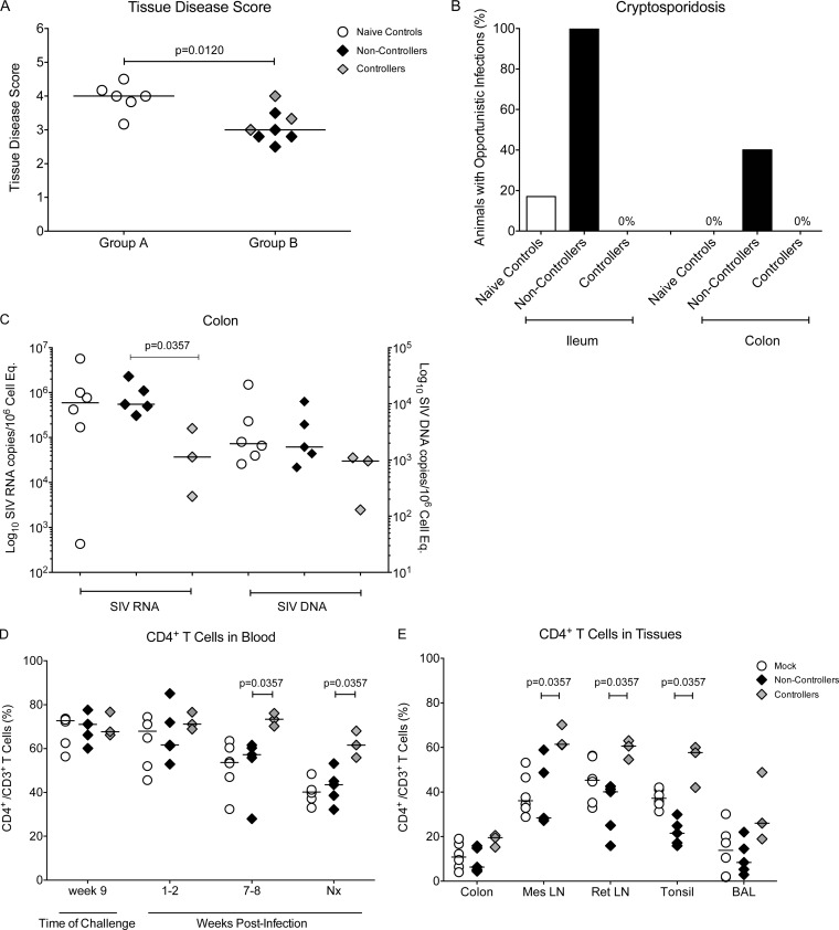
FIG 8.
Clinical assessment of SIV-infected animals. (A) The median disease score, determined by histological analysis, for animals in group A (naive controls) and group B animals at the time of euthanasia. Controller and noncontroller animals are represented by gray and black symbols, respectively. (B) The percentage of animals with detectable cryptosporidiosis in the colon and ileum of naive controls and vaccinated animals at the time of euthanasia. (C) SIV RNA (left y axis) and DNA levels (right y axis) were measured in colon tissue samples by PCR and are reported as copy numbers per 106 cell equivalents. (D) The percentage of CD4+ T cells within the CD3 population at the time of oral SIV challenge initiation (week 9), at weeks 1 to 2 (acute phase) and weeks 7 to 8 (chronic phase), and at the time of euthanasia in nonvaccinated animals and in controller and noncontroller vaccinated infant macaques. Median values per group are represented by horizontal bars. (E) Comparative analysis of CD4+ T cell frequencies in selected tissues of mock-vaccinated and vaccinated animals at the time of euthanasia. Differences between animals in 2 different groups were determined using the nonparametric Mann-Whitney test. Mes, mesenteric; Ret, retropharyngeal; Nx, necropsy.

