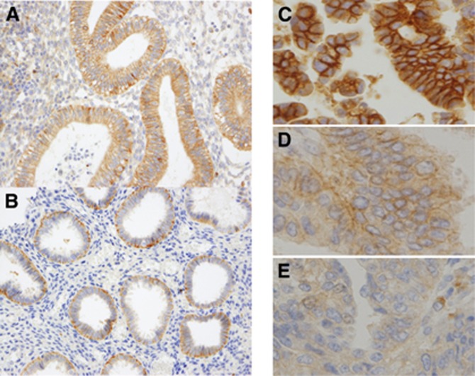
Figure 1.
Immunohistochemical analysis of DLG1 expression in normal and malignant endometrial tissues. (A) Expression of DLG1 in normal endometrium in the proliferative phase. (B) Expression of DLG1 in normal endometrium in the secretory phase. Human DLG1 expression was more abundant in the lower part of the basolateral membrane in endometrial tissues. (C) Strong membrane-bound expression of DLG1 in endometrial cancer (hDlg score 2). (D) Membrane-bound weak DLG1 expression in endometrial cancer (hDlg score 1). (E) Weak cytoplasmic DLG1 expression in endometrial cancer (hDlg score 0).

