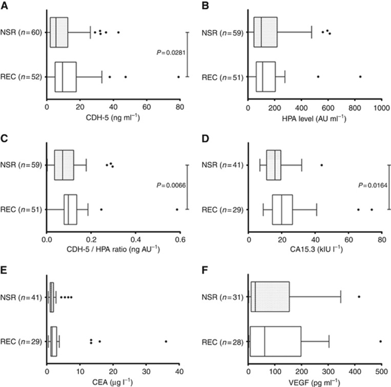
Figure 2.
Box plots showing biomarker levels in patients with either NSR or REC. (A) CDH; (B) HPA binding to (antibody) captured CDH; (C) Ratio of CDH5:HPA; (D) CA15.3; (E) CEA; (F) VEGF. The whiskers represent the data within 1.5 IQR of the upper and lower quartiles. Outliers are indicated by dots. Statistical significance indicated.

