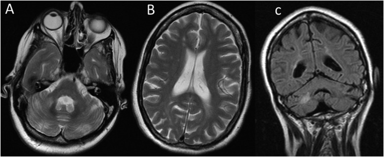
Figure 7.
Axial T2 (a, b) and coronal fluid attenuated inversion recovery (c) in female patient with FA. There is MRI evidence of cerebral and cerebellar atrophy with associated cerebral and cerebellar white matter signal changes in keeping with microvascular angiopathy beyond what would be expected in a patient of 47 years with no cardiovascular risk factors.

