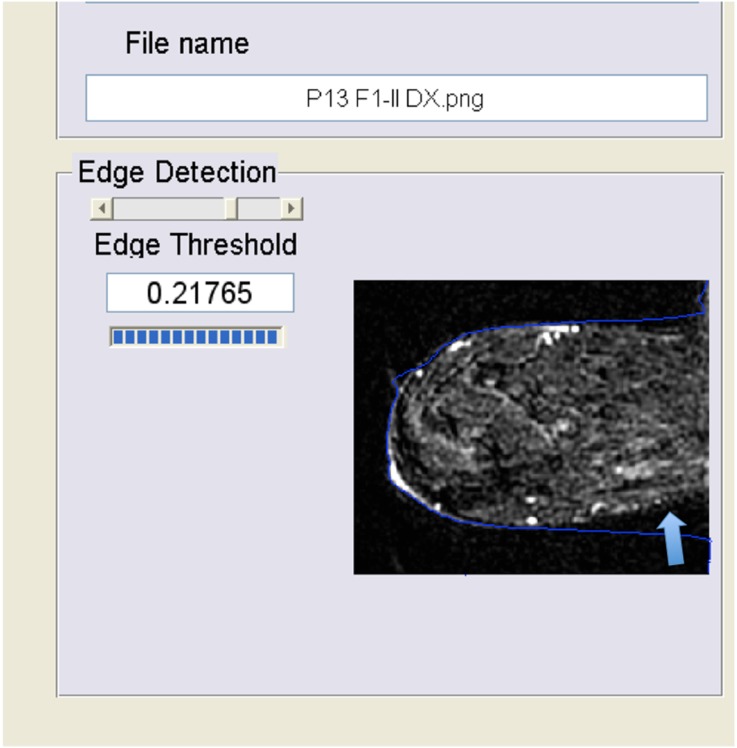
Figure 2.
Error in the edge detection near the pectoral region. In this case (error near the thoracic wall, arrow), the values of background parenchymal enhancement were underestimated by the automatic software owing to the inclusion of more background tissues. These data were corrected using the semi-automatic version of the software manually adjusting the edges.

