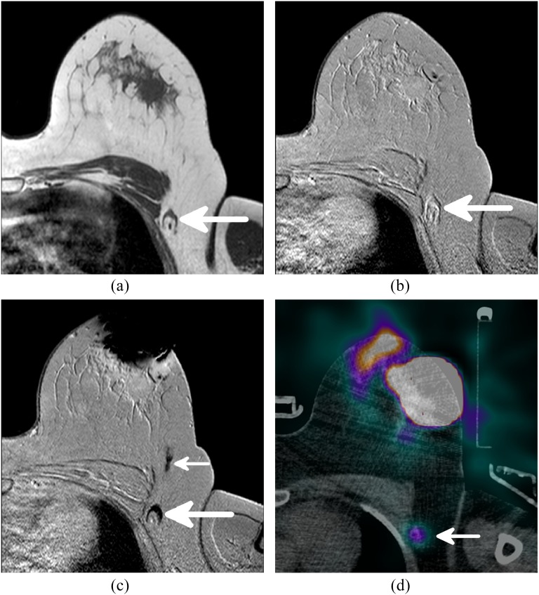
Figure 1.
The different MR sequences and single photon emission CT (SPECT)-CT of a single patient. (a) A T1 weighted image showing the anatomy and morphology, one axillary lymph node (LN) is visible (arrow). (b) The same axillary LN, visible (arrow), on a pre-contrast T2 weighted scan. (c) A large decrease in signal is observed at the periareolar injection site. The axillary LN also shows a signal decrease owing to uptake of superparamagnetic iron oxide (SPIO) and is therefore considered sentinel lymph node (SLN) (large arrow). A SPIO-filled lymphatic vessel draining from the injection site to the SLN is indicated with the small arrow. (d) The radioactive SLN (large arrow) identified by SPECT-CT corresponds to the SLN identified by MRI.

