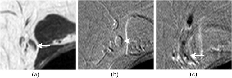
Figure 3.
A sentinel lymph node (SLN) showing inhomogeneous superparamagnetic iron oxide uptake. The arrow indicates the SLN on (a) T1 weighted turbo spin echo image, (b) pre-contrast T2 weighted gradient echo (GRE) image and (c) post-contrast T2 weighted GRE image. The position of the lymph node is different in the post-contrast scan owing to the movement of the patient during the procedure.

