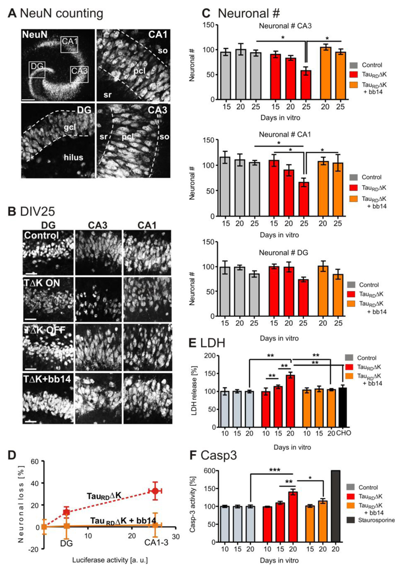
Fig. 6. Expression of TauRDΔK causes neuronal loss via activation of caspase 3.
(A) NeuN positive cell bodies were counted in the DG and within the pyramidal cell layer in area CA1 and CA3 (dashed lines, slice at DIV25).
(B) Representative neuronal cell layers from slice cultures at DIV25: control, TauRDΔK slices in switched-ON mode TauRDΔK slices in switched-off mode (DOX treated for DIV1-DIV25) and TauRDΔK slices in switched-on mode in the presence of compound bb14 (treated for DIV1-DIV25).
(C) Neurons in area CA1, CA3 and DG were counted at DIV15, 20 and 25 (8-10 slices, 6 animals/group). The number of neurons was strongly reduced in TauRDΔK slices, both within area CA1 (-28%; F(2/30)=4.405; p<0.05) and CA3 (-37%; F(2/31)= 5.410; p <0.05) at DIV25 in comparison to control slices. This effect was already observed at DIV20 (CA1 (-18%) and CA3 (-16%) (not significant) and less prominently at DIV15 (CA1 (-6%) and CA3 (-5%). The neuronal number of the DG was only slightly reduced at DIV25 (-14%) and unaffected at earlier time points. Neuronal loss could be prevented by compound bb14 (15µM, starting at DIV1) in all the subregions (e.g. CA3 (only 3% (DIV20) and 7% (DIV25)).
(D) Correlation between TauRDΔK expression (determined by reporter gene activity) and neuronal loss. The high expression of TauRDΔK in the pyramidal cell layer causes a strong neuronal loss, but not in the DG where the expression level was much lower (Pearson r = 0.9759, linear regression test).
(E) LDH release (cytotoxicity) was analyzed in TauRDΔK, TauRDΔK+bb14, TauRDΔK+DEVD-CHO and control slices at 10, 15 and 20 DIV. A pronounced increase (~40%; F(3/19)=7.736; p<0.001) in LDH release was observed due to TauRDΔK expression at 20 DIV but not at 10 DIV. At DIV15 the LDH release was slightly increased by 13% in TauRDΔK expressing slices. The presence of bb14 rescued the effect on LDH release at 20 DIV to control levels. Treatment of slice cultures with the caspase-3 inhibitor DEVD-CHO strongly prevents LDH release when comparing TauRDΔK, TauRDΔK+bb14, TauRDΔK+DEVD-CHO and control slices.
(F) Caspase 3 activity was analyzed in TauRDΔK, TauRDΔK+bb14 and control slices at DIV 10, 15 and 20. Caspase 3 activity was unchanged at DIV10. TauRDΔK expression caused an increase in caspase 3 activity by 10% (DIV15) and a significant increase by 35% (F(2/16)=12.20; p<0.001) (DIV20), compared with slices from control mice. Treatment with bb14 relieved apoptosis (p<0.05). Apoptosis inducer staurosporine (0.5 µM for 24 hours) increased the caspase 3 activity by ~600%.
One-way ANOVA followed by Tukey’s post-hoc test: *p < 0.05; **p < 0.01 and ***p < 0.001. Scale bars: A, upper panel, 200µm; A, lower panel, B, 50µM.

