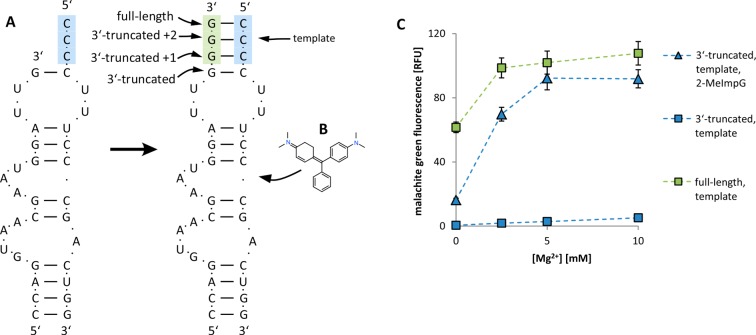
Figure 2.
Malachite green aptamer. (A) Structure and sequence of the malachite green aptamer before and after primer extension. Blue bases, template region; green bases, primer extension region. (B) One molecule of malachite green binds to each fully assembled aptamer. (C) Fluorescence of malachite green in the presence of aptamer RNA at different concentrations of Mg2+. Blue squares, unextended 3′-truncated strand plus template strand; blue triangles, 3′-truncated strand plus template after 24 h of RNA primer extension with 2-MeImpG; green squares, 3′-truncated strand after 24 h extension with 2-MeImpG and template strand. Each sample contained 0.25 M Tris-HCl pH 8.0, 0.15 M NaCl, 2 μM malachite green, 1 μM each strand of the aptamer. Error bars indicate SEM, N = 3.

