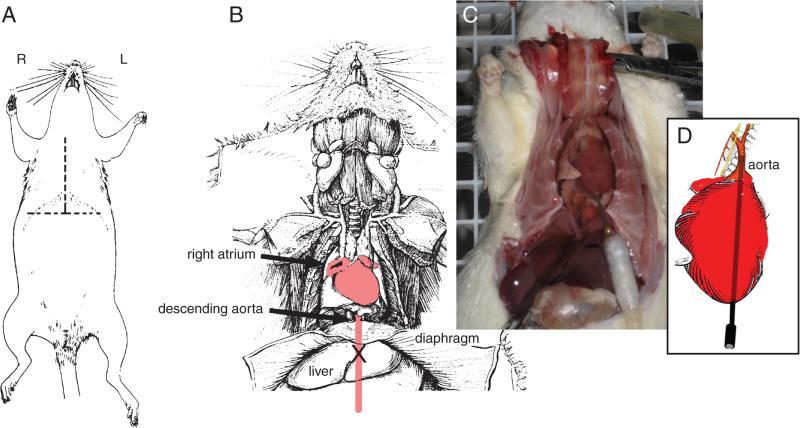
Figure 2.12.7.
The procedures used to perfuse a rat. The rat is anesthetized and an inverted “T”-shaped incision made (A). The diaphragm is cut and the rib cage opened to expose the heart (B and C). After injecting heparin into the heart at its apex, a catheter is inserted into the heart as shown in C. The tip of the catheter is eased into the proximal portion of the aorta as shown in D. The right atrium is cut (as illustrated in B to enable the perfusate to drain from the animal. (Photograph courtesy of Emmanuel Díaz, a student at the Ricardo Miledi Neuroscience Course, Universidad Nacional Autónoma de México, 2008).

