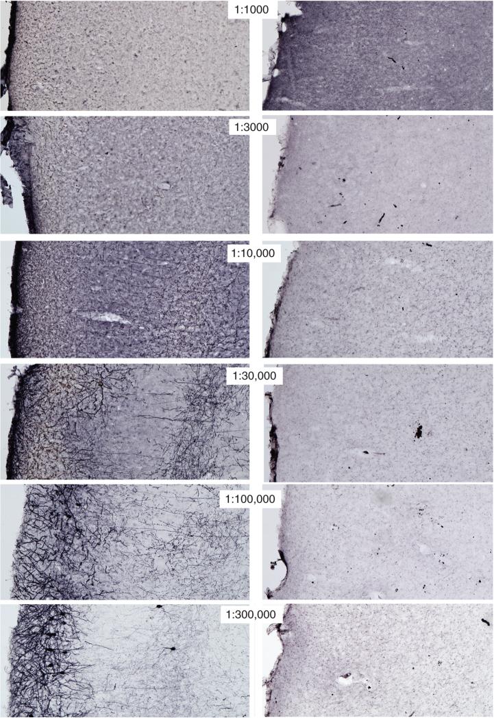
Figure 2.12.8.
Example of titrations of two purchased antisera (both rabbit polyclonal antisera) against green fluorescent protein (GFP). Both are tested in the same tissue. Note that the antiserum used in the series on the left (purchased from Novus) shows specific staining for GFP in neurons of the cortex, whereas only background labeling is found in the series on the right that used the other antiserum. For the color version of this figure go to http://www.currentprotocols.com.

