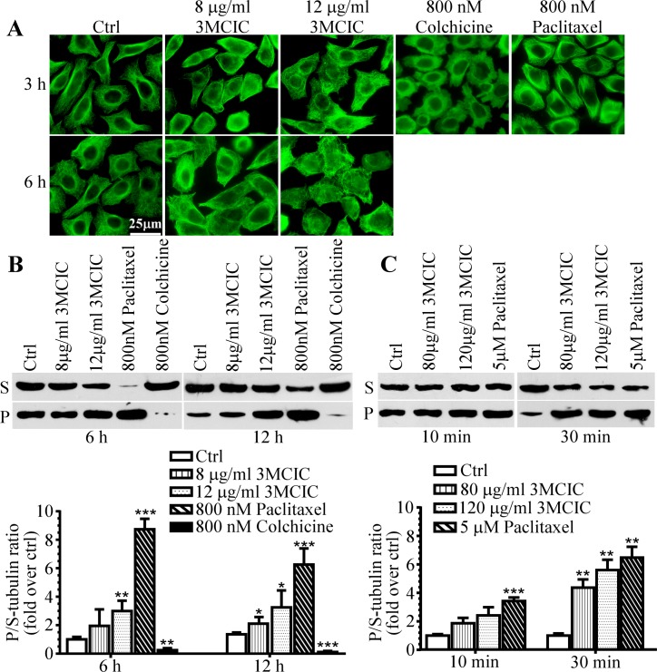
Fig 5. Influence of tubulin polymerization by 3MCIC.
HepG2 cells were incubated at 37°C with 3MCIC, paclitaxel or colchicine, and assayed with α-tubulin mAb by immunofluorescence (A) and Western blotting (B) after separation of soluble (supernatant, S) and polymerized (pellet, P) tubulins. (C) HepG2 cell lysates were incubated at 25°C for 10 or 30 min with 3MCIC or paclitaxel respectively, and analyzed by Western blotting. In both B & C, normalized changes of the band-intensity ratios of P/S-tubulin in drug-treated groups over controls were plotted. *P<0.05, **P<0.01 and ***P<0.001 vs control (n = 3).

