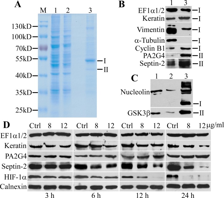
Fig 9. Target isolation by LAC and changes of target proteins in 3MCIC-treated cells.
HepG2 cell lysates were loaded onto the LAC column, and the eluted proteins were analyzed by SDS-PAGE (A) and Western blotting (B & C). The positions of band I and II are indicated at the right side. M is molecular weight markers. Lane 1: HepG2 cell lysates (20 μg total protein loading). Lane 2: a flow-through fraction from the column (15 μg for A and 20 μg for C). Lane 3: the column eluent (5 μg). (D) Regulation of target proteins in 3MCIC-treated HepG2 cells.

