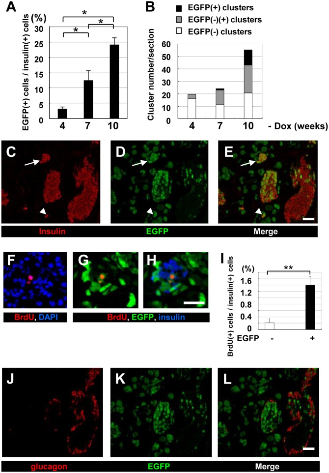
Fig 4. Immunohistochemical analysis of the pancreas of ERTF-Pdx1-EGFP mice maintained without Dox.
Sections from the pancreas of ERTF-Pdx1-EGFP mice 4, 7, and 10 weeks after Dox withdrawal (n = 3, 3, and 4, respectively) were stained for insulin, EGFP, and DNA. (A) The percentage of EGFP-positive cells among insulin-positive islet cells was calculated for each mouse. The total number of insulin-positive cells examined was 4968, 6690, and 9555 at 4, 7, and 10 weeks after Dox withdrawal, respectively. Statistical analysis was performed by one-way ANOVA followed by Tukey’s post-hoc test. *P < 0.05. (B) EGFP expression in islet-like clusters containing fewer than 10 insulin-positive cells was examined for the pancreas of these mice. The percentage of islet-like clusters containing insulin-producing cells with only EGFP-negative (open bars), EGFP-positive and negative (grey bars), and only EGFP-positive (black bars) cells was determined. (C-E) Immunohistochemical analysis of the pancreas of ERTF-Pdx1-EGFP mice 10 weeks after Dox withdrawal. Sections were stained for insulin (red) and EGFP (green). Right panel (E) is a merged view of (C) and (D). Arrow shows an islet-like cluster in which all the insulin-positive cells were also EGFP-positive. Arrowhead shows EGFP-positive insulin-producing cells in an islet-like cluster. Bar = 50 μm. (F-I) Increased BrdU-positive cells in the islets of ERTF-Pdx1-EGFP mice 6 weeks after Dox withdrawal. Pancreas sections were stained for BrdU (red), DNA (blue), and EGFP (green). Merged image of (G) with insulin staining (blue) is shown in (H). Bar = 25 μm. The percentage of BrdU-positive cells among EGFP-negative and -positive insulin-positive islet cells was determined (I). In the islets, BrdU-positive cells were significantly more enriched in EGFP/insulin double-positive cells than in insulin-positive EGFP-negative cells. The total number of insulin-positive islet cells examined was 1514, 1762, 2233, and 3375 each from four ERTF-Pdx1-EGFP mice. **P < 0.01. (J-L) Pancreas sections from ERTF-Pdx1-EGFP mice 10 weeks after Dox withdrawal were stained for glucagon (red) and EGFP (green). Serial section to that used in (C-E) was used. Right panel (L) is a merged view of (J) and (K). No EGFP/glucagon double-positive cells were observed. Bar = 50 μm.

