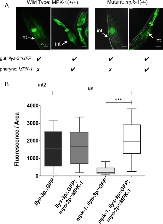
Fig 8. The activation of ILYS-3 is cell non-autonomous and requires MPK-1 activity in the pharynx.
(A) Images of single and double transgenic animals expressing ilys-3p::GFP only or in combination with the myo-2p::MPK-1 in mpk-1(ku1) and WT (N2) backgrounds. The myo-2p::MPK-1 construct drives MPK-1 expression in the pharynx and restored ilys-3 expression in the intestine (int) of mpk-1 mutants. (B) Quantification of fluorescence intensity (after background subtraction) in the intestinal cell int2 of single and double transgene reporter strains. Data analyzed with Mann–Whitney Unpaired test, 95% confidence level. Fluorescence intensity values for mpk-1(ku1); ilys-3p::GFP; myo-2p::MPK-1 vs ilys-3p::GFP; myo-2p::MPK-1 and mpk-1(ku1); ilys-3p::GFP; myo-2p::MPK-1 vs ilys-3p::GFP were not significantly different (NS). Mean values for mpk-1 mutants with the double transgene differ significantly from their sibling controls harbouring the ilys-3p::GFP reporter only (*** p = 0.0003). N = 10-15/group.

