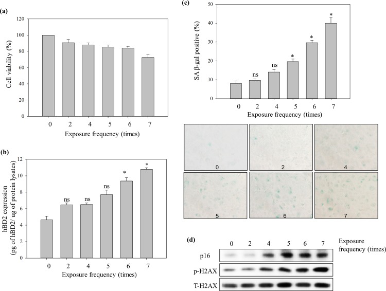
Fig 2. Repeated low energy UVB irradiation-induced increase of SA markers and hBD2 expression in NHEK.
NHEK were treated with repeated UVB radiation exposures (5 mJ/cm2) at scheduled exposure times (0 to 7 times) and the time interval between exposures was 30 min. (a) Viability, (b) induction of hBD2 expression, (c) β-gal activity and (d) SA-protein markers at 64 h after each repeated exposure to UVB on NHEK. The degree of cell senescence was quantified as the percentage of SA-β-gal positive cells and expressed as a percentage of the 0 exposure group. hBD2 expression was measured using whole cell lysates by ELISA assay. Data are presented as the mean ± SEM of three independent experiments (n = 3). *, p<0.05, control vs. UVB treatment group. ns, no significance.

