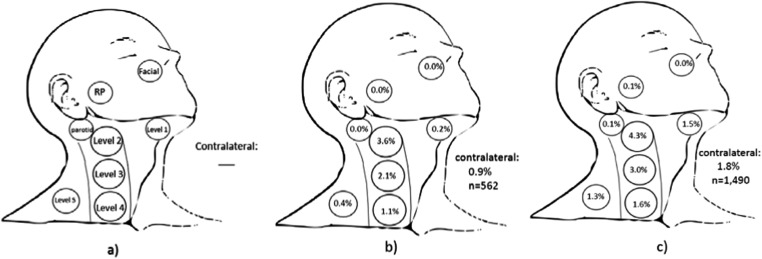
Figure 1.
Nodal metastases at presentation: (a) depiction of each of Levels 1–5, retropharyngeal (RP), intraparotid and facial nodes. Reference standard pictorial representation of risk of nodal involvement in patients with T2 glottic squamous cell carcinoma (b) with impaired mobility only (7.1% nodal involvement, n = 562) used for statistical comparison and (c) with subglottic or supraglottic extension (9.7% nodal involvement, n = 1490).

