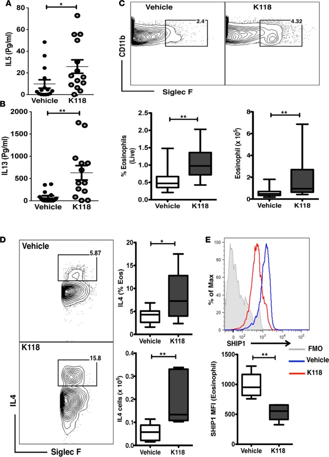
Figure 3. K118 treatment increases eosinophils in the visceral adipose tissue of DIO mice.
(A and B) 14- to 16-week-old diet-induced obese (DIO) mice treated with K118 (10 mg/kg body weight) or water (vehicle) for 2 weeks (twice per week), followed by determination of IL-5 and IL-13 cytokines serum levels (pg/ml) by ELISA (n = 13–15). (C) Flow cytometry plots showing gating for eosinophils and frequency (total viable epidydimal white adipose tissue [eWAT]) and number of eosinophils in the eWAT of K118- and vehicle-treated mice (n = 15). (D) Flow cytometry plots showing analysis IL-4–expressing SiglecF+ eosinophils, their frequencies (percentage of eosinophils), and numbers in the eWAT (n = 10). (E) Expression of SHIP1 in the eosinophils of eWAT of DIO mice (n = 5) treated for 2 weeks with K118 (red) or vehicle (blue). Student’s t test,*P < 0.05,**P < 0.01. Data are represented as mean ± SEM. Sample sizes are biological replicates. Box-and-whisker plots are defined as follows: the bounds of the boxes indicate SD; the lines within the boxes indicate means, and the whiskers represent minimum and maximum values.

